Schistosomiasis, also known as water belly or snail disease, is an infection caused by parasites of the genus Schistosoma mansoni (S. mansoni). Schistosoma species are responsible for the disease in the Americas.
Just summarizing what was explained in detail in the first part, the Schistosomiasis is a disease caused by a parasite that inhabits the blood vessels of the intestinal system. There it releases thousands of eggs that are shed in the feces. The embryo contained in these eggs, called miracidium, needs water to be free and the snail to multiply. Large freshwater reserves, such as lakes and reservoirs, which are inhabited by snails, are the ideal locations for the spread of schistosomiasis. After multiplication inside the snail the miracidium becomes a larva, called cercaria, and returns to water. Anyone who bathe or drink water contaminated with cercariae can become infected. The larvae penetrate into the skin, blood vessels and reach the vessels of the liver and intestines, where they lay their eggs, restarting the cycle.
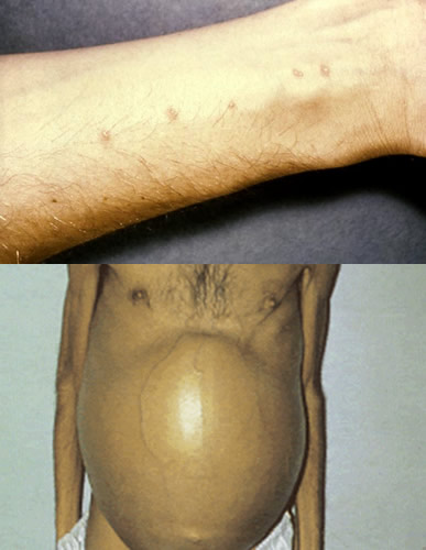
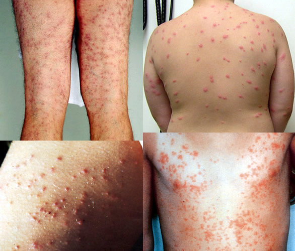
The first symptoms of schistosomiasis may occur immediately after the invasion of the skin by the parasite. The penetration of the skin by the cercariae may cause so-called "swimmer's itch". This is one or more lesions, typically in the legs or feet in the form of red papules that cause intense itching.
Not all people infected by cercariae have this skin lesion. When it occurs, an immediate tingling and itching at the site of entry may occur, followed by a pruritic papular eruption that appears within 12-24 hours, and can last over a week.
Importantly, the swimmer's itch can also be caused by other species of Schistosomas, which infect birds and other mammals, not being able to infect humans. In these cases the cercariae invade the skin but is eliminated by the body after a few hours without causing major consequences. On the American continent only S.mansoni cercariae are able to survive and cause schistosomiasis in humans.
Most people living in endemic regions of S.mansoni are contaminated in infancy and remain with the parasite in their digestive system silently for many years. Sometimes the initial symptoms are minor and some end up being confused with common childhood illnesses.
Katayama fever
Fever
The incubation period of schistosomiasis, i.e. the interval between the contamination and the first symptoms of the disease itself, is from one to two months, which corresponds to the phase of penetration of cercariae, their maturation to the adult form and setting of S . mansoni in blood vessels.
The Katayama fever is acute schistosomiasis, caused by a reaction of the immune system due to migration and production of parasite eggs in the body. It occurs between two to eight weeks after exposure. The acute phase usually arises in the community outside endemic areas and without previous contact with the parasite. Individuals living in endemic areas and have been exposed to S. mansoni during childhood often have no early symptoms.
Katayama fever symptoms include fever, chills, muscle pain, joint pain, dry cough, diarrhea, loss of appetite and headache. During the physical examination swollen lymph nodes (glands) and hepatosplenomegaly (swelling of the liver and spleen) can be found. Symptoms usually resolve spontaneously over a period of several weeks. In rare cases, if there is a massive invasion of parasites and the immune reaction is severe, the patient may progress to death.
Chronic infection by schistosomiasis
Contrary to what occurs in the acute phase, the complications related to chronic schistosomiasis are more common in endemic areas, where individuals are at increased risk of a high parasite load and frequent contamination. However, it is important to emphasize that not all patients infected with the parasite develop symptoms of chronic schistosomiasis. Some patients remain contaminated by eliminating eggs in the feces, but show no signs of disease.
The chronic form starts from the sixth month after infection and can last for several years. The severity of the chronic form is related to the quantity of parasites and location where they lay their eggs.
The Schistosoma are ususally located in mesenteric veins, which are the blood vessels that drain the intestines. The mesenteric veins drain blood toward the door where a large vein is that receives all the blood from the digestive system and leads to the liver.
The parasite eggs are generally deposited in mesenteric veins, but both may migrate into the intestine as the portal vein and liver.
The chronic schistosomiasis, which is much more common than the acute form of the infection, is caused by the body's immune response to eggs, resulting in severe inflammation of the tissues affected and progression to fibrosis and granulomas (replacement of normal tissue with scar tissue).
The chronic form of schistosomiasis has the following forms:
The retention of eggs in the intestinal wall causes bloody diarrhea, cramps and weight loss. The intense inflammatory response of the body against the eggs may cause ulcers in the gut wall, granulomas and obstruction to the passage of stool.
Patients infected with a large load of parasites are more likely to produce disease in the liver. The eggs of the parasite tend to migrate and settle in the portal vein, causing inflammation and obstruction of the passage of blood by fibrosis.
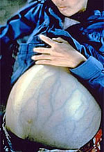
As all the blood coming from gastrointestinal system passes through the portal vein to the liver before going to the rest of the body, an obstruction in this region causes a huge "traffic jam" of blood, which leads to what we call portal hypertension. If no blood reaches the liver, it has to find other ways to get to the rest of the body, forming a collateral circulation.
Portal hypertension is responsible for complications of hepatosplenic schistosomiasis, among them, ascites, splenomegaly (enlarged spleen) and esophageal varices. The esophageal varices are a feared complication of portal hypertension, as they may rupture causing severe gastrointestinal bleeding and bloody vomiting.
Many patients infected by S. mansoni remain asymptomatic, so the suspicion of infection often arises by chance through routine blood tests. CBC can detect anemia and increased eosinophils, which speak in favor of a parasitic infection.
Diagnosis is made by stool testing, which is capable of detecting the eggs of Schistosoma. The parasite is usually only detected after six weeks of egg contamination. If the suspected schistosomiasis is large, but the stool test is negative, biopsy of the rectum (the final portion of the intestine just before the anus) can detect eggs.
In patients with clinical signs of portal hypertension, an ultrasound may be useful in identifying fibrosis in the portal vein caused by the deposition of eggs of Schistosoma.
Specific blood tests against S. mansoni are under development but not yet available to the public.
The Praziquantel is the drug against schistosomiasis. The recommended dose is 60 mg/kg for children up to 15 years and 50 mg/kg for adults, both in a single dose. The medication is presented in 600 mg tablets, divisible into two equal parts in order to facilitate adjustment of the dose.
An alternative to the Praziquantel Oxamniquine is recommended at a dose of 15 mg/kg for adults and 20 mg/kg for children up to 15 years, both also in a single dose. There are two presentations: 250 mg capsules and suspension containing 50 mg per ml.