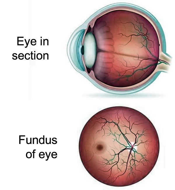
The fundus of the eye is the richest and most detailed live painting and color of the situation of the arteries, veins and nerves of the human body, since in its visualization only transparent means interposes between the physician and the retina of the patient, except in pathological situations. The retina works as well as a window through which one can see the health of the organism in general.
The retina is a nerve tissue sensitive to light, located on the inner surface of the back of the eye, whose function is to transform the light stimulus into a nerve stimulus. Compared with the photographic process, the retina is like the machine's film, which captures images through its photoreceptor cells to send them to the brain.
The fundus or ophthalmoscopy is the exam that you visualize the fundus structures, paying attention to the optic nerve, retinal vessels and the retina itself, especially its central region called the macula. The eye examination has been carried out since 1851, when von Helmholtz invented the first ophthalmoscope and is the main link between ophthalmology and other branches of medicine. The optical principle consists of the projection of light from the ophthalmoscope inside the eye and through the reflection of that light in the retina it is possible to observe these structures.
There are two types of fundoscopy: direct, in which an enlarged image is obtained fifteen times larger, but with restricted field of vision, and the indirect, which provides an image with smaller enlargement, but with a wider visualization of the retina, to its periphery. Direct ophthalmoscopy is usually performed by the general practitioner and a simple and portable device is used, while indirect ophthalmoscopy is usually restricted to the ophthalmologist and depends on more complex equipment.
Individuals who do not have eye problems or diseases that predispose them to eye diseases, such as high blood pressure and diabetes, need to be tested annually, especially if they are over 40. The general practitioner or ophthalmologist will suggest a different periodicity for those who are following up for an eye or systemic disease, taking into account the patient's history.
The fundus examination can bring important information to individuals of all ages. Preterm infants whose mothers had infections during pregnancy should be routinely screened for a detailed fundus examination. All other infants should be evaluated with the red reflex test, which evaluates the reddish coloration generated by the retina through the pupil when submitted to direct illumination. This test can be performed by the pediatrician himself, in the nursery or in the delivery room, who will refer him to the ophthalmologist in the event of any change or a dubious result. Ophthalmoscopy, in these cases, may indicate the presence of tumors such as retinoblastoma, infections such as toxoplasmosis, rubella, cytomegalovirus and syphilis, as well as diseases such as retinopathy of prematurity.
In adults, regular eye examination is essential for the early diagnosis of various eye diseases, including glaucoma. Allied to the measurement of intraocular pressure, it allows treatment to be started before the presence of symptoms, since glaucoma, the second cause of blindness, is a silent disease that can take years to cause visual difficulty.
Age-related changes, such as the appearance of drusen in the retina and the development of age-related macular degeneration, are also always observed by the ophthalmologist during fundoscopy.
Fundoscopy, due to its links with the medical clinic, neurology and other specialties, is an important element for the diagnosis and follow-up of various systemic diseases. In subjects with hypertension or diabetes careful funduscopy can bring valuable information about the underlying vascular situation. It is interesting to note that fundoscopy is a practical and easy method to evaluate the damages in target organs, besides providing information about the activity and time of development of these diseases. This examination is one more means to be used by the clinician for a better monitoring and treatment of these diseases and to prevent their ocular and systemic complications.
In neurology, fundoscopy is commonly used, for example, in coma patients in search of signs such as papillary edema that may indicate intracranial hypertension and the presence of subhyaloid hemorrhage, which may suggest intracranial hemorrhage.
Thus, in recent decades, where we have witnessed great advances in medicine, prevention naturally assumes a fundamental role in promoting health. The wealth of information contained in the eye fundus is a valuable tool in this patient care.