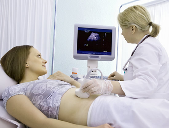
In pregnant women, tests that use radiation, such as X-rays and computed tomography, should be avoided due to the obvious risks that they bring the baby. Therefore, ultrasound is the imaging method of choice for prenatal care. Ultrasound is an inexpensive test without risk to the fetus, it causes no side effects and has no contraindications.
In this article we will make a brief review of the role of obstetric ultrasound. We will address topics such as nuchal translucency, morphological ultrasound estimated gestational age and 3D and 4D ultrasound.
Ultrasonography can be performed repeatedly during pregnancy, but it is not needed in every query. In fact, many international societies of Obstetrics and Gynecology agree that if the patient is healthy, has no complaints or risk factors, only one or two sonographic evaluations were carried out between the 10th and 13th week and between the 16th and 20th weeks of pregnancy they are really necessary throughout pregnancy.
But like many obstetricians have an ultrasound device in your office, it is very common that the examination is over part of many, if not all, of the prenatal visits. It's good to make it clear that being pregnant as well, there are no studies that indicate benefits in terms of health of the mother and the fetus when compared pregnant women who received ultrasound series throughout pregnancy with pregnant women who received only a single ultrasound between the 16th and 20th week of pregnancy. Therefore, in places with structure and limited access to medical resources, there is nothing wrong in asking just a single ultrasound during pregnancy.
In places with more resources, the obstetrician usually hold at least 3 or 4 ultrasound examinations in the prenatal, divided over the three trimesters of pregnancy:
1. In the first or second prenatal visit, with the objective of confirming the existence of an embryo within the gestational sac and intrauterine pregnancy, visualizing the heartbeat of the fetus early to identify a twin pregnancy, estimate gestational age and evaluate possible abnormalities of the female gynecologic tract, such as ovarian cysts, fibroids, uterine malformations, etc.
2. In the second trimester, ultrasonography should be performed, preferably between the 18th and 24th week of gestation to assess the anatomical formation of the fetus. This examination is called morphological ultrasound (or morphological ultrasound) and is the most important ultrasound pregnancy, it is able to detect fetal malformations.
Early in the second quarter, around the 12th week of pregnancy, it is also very common to have an ultrasound to measure nuchal translucency, which is a test that measures the amount of fluid in the fetal nuchal region. A nuchal translucency suggests the possibility of a chromosomal alteration, such as Down's syndrome.
3. In the third quarter ultrasound is used to monitor the growth rate of the fetus, the placenta location of the uterus, the amount amniotic fluid, fetal vitality, its position within the uterus and the position of the umbilical cord.
The obstetrical ultrasound in the first 12 weeks of pregnancy are usually made by transvaginal. From the 12th week, the abdominal approach is the most appropriate.
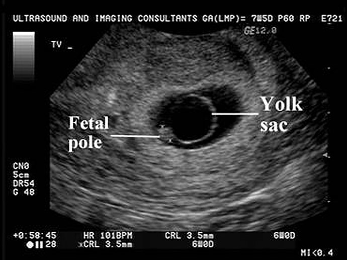
Yolk sac in ultrasound
The first ultrasound in pregnancy can be performed as early as the 5th week of pregnancy, a time when already can see the gestational sac, first identifiable structure of a pregnancy. The gestational sac tends to be visible from 4.5 weeks of gestation. Before the 4th week no point doing ultrasound, because it is not able to identify any signs of pregnancy.
Some days after the beginning of the 5th week of gestation is possible to identify, within the gestational sac, the yolk sac structure that provides nutrients to the embryo. The presence of gestational sac and yolk sac within the uterus confirms the existence of an intra-uterine pregnancy, discarding the possibility of an ectopic pregnancy, even if the embryo can not be viewed.
The embryo itself is usually visible from the 6th week and your heart rate can be identified between the 6th and the 7th week.
In general, we indicate the completion of the first ultrasound during the 7th week of pregnancy, when all the data mentioned above will be available to the obstetrician.
In the first trimester of gestation, the human embryo has a rate of growth and development more or less stable and predictable, and thus possible to estimate the gestational age according to its sonographic features. From the 2nd quarter, babies begin to grow at different speeds, according to their genetic characteristics and pregnancy conditions, more difficult to estimate the gestational age at ultrasound.
The estimated gestational age and expected delivery date (DPP) are made more accurately by obstetrical ultrasound in the first quarter of the last menstrual period (LMP), especially in women who have an irregular menstrual cycle or not remember with certainty the starting day of the last menstrual period. If the DPP calculated by LMP is different from the DPP calculated by ultrasound, the latter should be used by obstetricians to assess the most accurate gestational age.
The size of the gestational sac and mainly a measure called crown-rump length (CRL) are the measurements commonly used to estimate gestational age. Data, such as the presence of heartbeat, the yolk sac and a visible embryo inside the uterus also help in estimating the gestational age in pregnancies still very early.
From the 12th week of pregnancy, other measurements can be used to estimate gestational age, such as biparietal diameter (BPD), head circumference (WC) and femur length (CF).
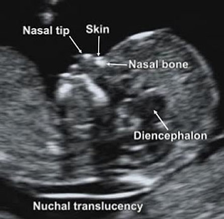
The assessment of nuchal translucency (NT), which some people call morphological the first quarter ultrasound is an ultrasound examination usually performed between the 11th and 13th week of pregnancy, which aims to identify the amount of liquid present in the neck of the fetus. Studies show that fetuses with chromosomal abnormalities, fetal malformations or genetic syndromes tend to have more fluid in this region, making the neck look wider.
Nuchal translucency
As liquid form fewer echo solid structures, ultrasound, they appear more darkened, or more translucent or nuchal translucency. The examination of nuchal translucency has value only if it is performed in fetuses with crown-rump length between 45 and 84 mm and less than 14 weeks of gestation.
Normal values of nuchal translucency are smaller than 2.5 mm. These values, however, should be evaluated according to the age of the pregnant woman. A nuchal translucency greater than 2.5 mm in a pregnant woman of 22 years is less worrying than the same value in a pregnant 40 years. The higher the value of nuchal translucency, the greater the chance of the fetus have any genetic alteration.
The nuchal translucency can be an indicator of various diseases or genetic abnormalities, Down syndrome is the most important. About 75% of babies with Down syndrome have increased nuchal translucency.
It is necessary to strengthen the examination of nuchal translucency is just a screening test, not intended as a definitive diagnosis for nothing. The false positive rate is relatively high, around 5%. Similarly, a normal TN, below 2.5 mm do not rule out the possibility of a chromosomal disorder, for about 20 to 25% of fetuses with Down show normal NT.
When we find an increased nuchal translucency, other sonographic evaluations should be performed to obtain more data. Generally, the absence of nasal bone and blood flow changes in the ductus venosus (communication between the umbilical vein and inferior vena cava of the fetus) are alterations that enhance the possibility of genetic disorders.
When screening for nuchal translucency, nasal bone or ductus venosus are changed, an investigation with maternal blood analysis it is necessary (beta hCG and Plasma Protein (PAPP-A)).
With the data of TN and the results of blood tests, the obstetrician can calculate the risk of chromosomal diseases of the fetus. If the value is much higher than expected for age, amniocentesis (amniotic fluid collection) is usually indicated for definitive diagnosis.
The sex of the baby can now be identified by ultrasound from the 11th week of pregnancy. At this stage however, the success rate is only 70%. The sex of the fetus can be detected more securely from the 13th week of pregnancy.
Fetal morphological ultrasound is the most important sonographic pregnancy. It should be done by cesarean section between 18th and 24th week of pregnancy. At this stage, the fetus is already fully formed, it being possible to identify relatively easily defects present.
The morphological ultrasound is the most time consuming and detailed pregnancy and can take more than half an hour, because the doctor needs to individually evaluate several different structures. In many cases, is not your obstetrician who performs this test, but a radiologist or other specialist obstetrician in fetal morphological ultrasonography.
In the morphological ultrasound can confirm the sex of the baby, check your heart and its chambers, evaluate the formation of the brain, the digestive organs, limbs, face and other systems of the fetus. The use of doppler serves to see how the blood flow in the placenta and uterus. This ultrasound is also possible to determine the location of the placenta, to see if it may be blocking the exit of the uterus, a condition called placenta previa.
The morphological ultrasound is also used to measure the baby's head, the femur and waist circumference to see if its growth is adequate.
Ultrasonography 3D has gained popularity in recent years due to increased clarity and beauty of its images. For parents, the 3D ultrasound is much more interesting because it shows the appearance of the fetus in much more detail. For the doctor, however, in most cases, there is no indication for its realization, because the 3D ultrasound adds little in relation to the common 2D ultrasound.
In some cases of suspected facial abnormality or neural tube seen the common ultrasound, 3D ultrasound appears to show defects with slightly higher sharpness. Out situations like this, there are few cases where the 3D ultrasound is really helpful.
4D ultrasound is only a 3D ultrasound seen in real time, able to show movements of the fetus and its internal structures such as the heart. It can be saved as a video, which makes it even more attractive to parents.
The image shown of the first trimester baby and the "yolk sac" is labeled incorrectly. The yolk sac should never be that big and the image is either misleading or there is a pathological issue